
Cancer cells grip and tear their way into new tissues
May 12, 2026Researchers from the National University of Singapore (NUS) have discovered that cancer cells do not simply push through surrounding tissues to spread, but instead actively grip onto protective tissue barriers and pull them apart, revealing a fundamentally new mechanism of cancer invasion that could open fresh avenues for therapeutic intervention.
Gripping, not pushing
Cancer cells can spread to distant tissues and organs, where they establish new tumours and ultimately lead to organ dysfunction and death. In ovarian cancer, clusters of tumour cells must break through a thin protective lining called the mesothelium, which covers the inner surface of the abdomen, in order to colonise new sites. These tumour clusters are typically thought to forcibly push their way through such tissue barriers in a process termed “invasion”. However, the cellular and molecular events driving this process have remained unclear.
Using laboratory-grown ovarian cancer cell clusters placed onto a mesothelial cell layer to mimic the invasion process, the research team led by Associate Professor LOW Boon Chuan, with research overseen and spearheaded by Dr Selwin WU, both from the NUS Department of Biological Sciences and NUS Mechanobiology Institute (MBI), discovered that instead of simply pushing through, both the interacting cancer cells and surrounding tissue behave in a manner akin to gripping with tiny claws, latching and pulling onto each other. This intercellular behaviour is mediated by integrin adhesion proteins, which transmit mechanical forces through these connections. Over time, this process causes the surrounding tissues interacting with the invading cancer cells to tighten and stretch, creating an opening that allows the invading cancer cells to spread and colonise new environments. This research work is a collaboration with Professor Yuan LIN from The University of Hong Kong.
The research breakthrough was published in the journal Developmental Cell.
Challenging the prevailing model
The prevalent explanation for cancer spread is that cancerous cells from tumours undergo a transition from stiff, solid-like forms to a more elastic, fluid-like state. This transformation allows cancer cells to push through tissues with ease. However, the new findings in this study change this understanding. Using advanced three-dimensional cell tracking and artificial intelligence-based analysis, the team showed that instead of switching between stiff and fluid states, cancer cells are already in a fluid-like state even before invasion begins, and that no such transition is required. Instead, the cancer cells interact and engage with the surrounding tissues by pulling at them, transmitting forces to these tissues, ultimately leading to tension build-up until the protective tissue gives way and tears apart.
The lead contact of the publication, Dr Selwin Wu, said, “This project builds directly on my previous research in USA and Australia exploring how tumour cells interact with their microenvironment. To uncover this mechanism, I brought together a multidisciplinary collaboration, integrating Associate Professor Low Boon Chuan’s leadership in biochemistry with Professor Yuan Lin’s expertise in materials engineering, alongside leaders in cell biology, pathology and physics. Driven by key experiments conducted together with Celestine Ho, our coordinated team was able to translate our interdisciplinary discoveries into a new model for future cancer therapeutics.”
Associate Professor Low said, “Our findings fundamentally change the way we think about cancer invasion. The tissue barrier is not merely a passive wall that cancer cells break down. It is mechanically involved in the process, responding to forces from the cancer cells in a way that ultimately leads to its own destruction.”
| A three-dimensional immunofluorescence-stained cancer spheroid binds to the protective tissue barrier, known as the mesothelium, beneath it. This interaction triggers tearing of the tissue barrier through combined forces from both the spheroid and the barrier. This study challenges prevailing paradigms of cells invading tissues. [Image credit: Celestine HO and Selwin WU] | 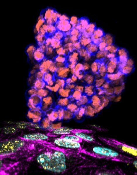 |
Implications for future therapies
These findings answer prevailing questions about the mechanisms by which cancer colonises new organs. Personalised medicine and therapeutic drugs can be designed to target the specific components of invading cancer cells and their target host cells that are involved in the sensing and transmission of these forces, potentially disrupting the pulling engagement or reducing the tension that leads to tearing of the protective tissue.
“Understanding these precise mechanical steps gives us concrete molecular targets to work with, bringing us closer to strategies that could prevent cancer from gaining a foothold in new organs,” said Associate Professor Low.
Reference
Wu SK*; Sun F; Ho CZ; Lou Y; Huang CBX; Nai MH; Xiao J; Shagirov M; Chin JFL; Lim D; Verma S; Tan DSP; Marcq P; Yap AS; Lim CT; Hiraiwa T; Lin Y*; Low BC*, “Multiscale mechanisms driving tissue rupture by invading cells” Developmental Cell DOI: 10.1016/j.devcel.2026.01.016 Published: 2026.
Footnote
The project is principally supported by the Singapore Ministry of Health’s National Medical Research Council under its Young Individual Research Grant (MOH-000653, Principal Investigator: Dr Selwin Wu). Other support includes funding from the Ministry of Education Tier 1 and the Singapore National Research Foundation (NRF) for the MBI mid-sized grant (NRF- MSG-2023-0001) to A/P Low.
Updated: 25 Jun 2026